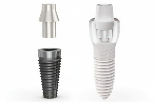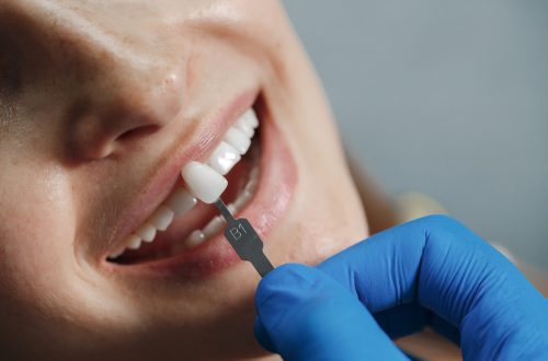
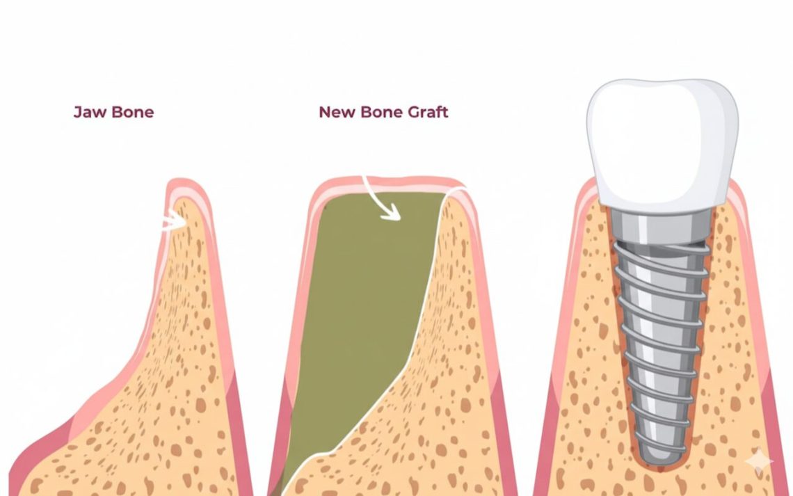
Dental implants are widely recognized as the gold standard for replacing missing teeth, offering unparalleled stability and functionality. However, the long-term success of an implant is entirely dependent on one critical factor: the health and volume of the underlying jawbone. This is where Bone Augmentation comes into play. Bone augmentation is a group of surgical procedures—including bone grafting and sinus lifts—designed to rebuild and restructure the jawbone where bone mass has been lost. These procedures are often a necessary prerequisite to ensure the implant fixture can be securely anchored in dense, native bone.
But why is augmentation so frequently required? Following the loss of a natural tooth, the surrounding alveolar bone begins to undergo atrophy (resorption). This occurs because the bone tissue no longer receives the functional stimulation previously provided by the tooth root during chewing. Without this biomechanical load, the body perceives the bone as unnecessary, leading to a progressive reduction in both height and width. This process can quickly leave the jawbone too shallow or too thin for standard implant placement, forcing the clinician to choose between complicated surgery or a traditional, less optimal prosthetic alternative. Therefore, bone augmentation procedures are essential tools that transform challenging anatomical sites into viable foundations for predictable, long-lasting implant therapy.
1. Understanding Bone Grafting (Augmentation)
Bone grafting, or bone augmentation, is a fundamental procedure in modern implantology. It involves using specialized materials to restore the dimensions of the alveolar ridge—the portion of the jawbone that supports the teeth. This intervention is often the difference between being ineligible for a fixed restoration and successfully receiving a long-lasting dental implant. The process aims to provide a structural scaffold that encourages the body to regenerate and replace the graft material with its own viable, load-bearing bone.
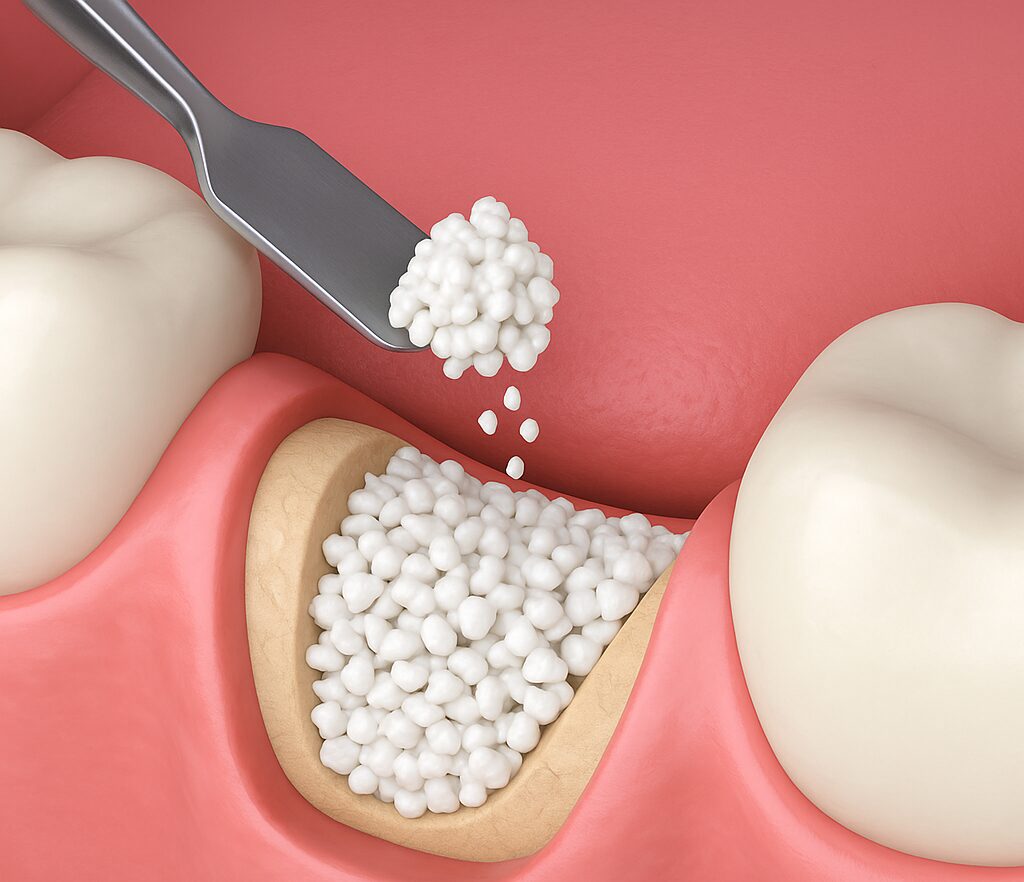
1.1. When is Grafting Required?
The necessity of bone grafting is meticulously determined during the initial planning phase using advanced imaging, primarily Cone-Beam Computed Tomography (CBCT) scans. Grafting is required when a patient presents with atrophy that has compromised the jawbone in three critical ways:
- Insufficient Vertical Height: The most common issue. Bone height is too low to fully embed the implant post without risking perforation of the maxillary sinus (in the upper jaw) or damage to the inferior alveolar nerve (in the lower jaw).
- Insufficient Horizontal Thickness (Ridge Width): The bony ridge has narrowed substantially, often after prolonged tooth loss, making the ridge too sharp or thin to provide the mechanical primary stability necessary for a successful implant placement.
- Aesthetic Deficiencies: Even if mechanically stable, bone loss can lead to gum recession, causing the final crown to look unnaturally long or lacking proper gum support. Grafting restores the contour required for optimal aesthetics.
1.2. The Four Types of Graft Materials
The successful outcome of the procedure depends heavily on selecting the appropriate graft material. These materials are classified based on their source, which determines their biological potential and handling characteristics:
- Autogenous Grafts (Autografts):
- Source: The patient’s own bone, typically harvested from the chin, jaw, or hip.
- Mechanism: This is considered the “Gold Standard” because it is the only material that is both Osteoinductive (contains living bone cells and growth factors) and Osteoconductive. This allows for the fastest and most reliable bone formation.
- Allogeneic Grafts (Allografts):
- Source: Human donor bone that has been carefully processed, freeze-dried, and sterilized to ensure safety.
- Mechanism: Primarily Osteoconductive, providing a matrix for regeneration. Its main clinical benefit is providing volume without the need for a second surgical harvesting site on the patient.
- Xenogeneic Grafts (Xenografts):
- Source: Non-human bone, most commonly derived from bovine (cow) or porcine (pig) origin.
- Mechanism: Strictly Osteoconductive. It acts as a long-lasting, stable scaffold due to its slow resorption rate. It is a very common choice for socket preservation.
- Alloplastic Grafts (Alloplasts):
- Source: Synthetic, biocompatible materials such as calcium phosphate or ceramic polymers.
- Mechanism: Strictly Osteoconductive. These materials are readily available, highly sterile, and used to fill small defects or maintain ridge height.
1.3. The Mechanism of Bone Regeneration
The function of a bone graft is defined by two key biological processes that guide healing and integration:
- Osteoinduction: This is an active process. The material contains or signals for growth factors and proteins that actively stimulate the surrounding host cells to differentiate into new bone-forming cells (osteoblasts). Autogenous grafts are the only materials that are inherently osteoinductive, which is why they offer the fastest and most reliable bone formation.
- Osteoconduction: This is a passive process where the graft material acts as a physical scaffold or matrix. This structure provides a secure latticework for the infiltration of blood vessels, osteoblasts (bone-forming cells), and other regenerative cells from the surrounding native bone. Almost all graft materials possess osteoconductive properties.
2. Sinus Lift Procedures (Upper Jaw)
While general bone grafting addresses deficiencies in height and width across the jaw, the Sinus Lift, or Sinus Augmentation, is a specific procedure reserved for rebuilding bone in the posterior region of the upper jaw (maxilla). This area presents a unique anatomical challenge due to its proximity to the maxillary sinus cavity.
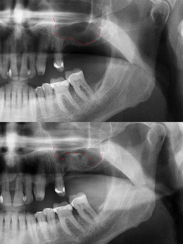
2.1. The Anatomical Challenge (Maxillary Sinus Pneumatization)
The need for a Sinus Lift arises from two simultaneous physiological processes following the loss of upper molar and premolar teeth:
- Alveolar Bone Resorption: The supportive bone recedes upwards due to the lack of mechanical stimulation from the tooth root.
- Maxillary Sinus Pneumatization: The floor of the maxillary sinus cavity often expands downwards into the space previously occupied by the tooth roots.
These two factors combined can reduce the available vertical bone height to just a few millimeters, making it impossible to place a standard implant without piercing the delicate Schneiderian membrane (the sinus lining) and entering the air-filled cavity. The goal of the Sinus Lift is to carefully elevate this membrane and fill the space with bone graft material, thereby creating a new, stable base for the implant.
2.2. Lateral Window Technique (Open Sinus Lift)
The Lateral Window Technique is the gold standard for repairing severe bone deficiencies in the posterior maxilla (typically less than 5 mm of native bone height). It is the most predictable method for gaining substantial vertical bone volume.
- Procedure: The surgeon accesses the sinus floor by creating a small, hinged “window” (or flap) in the lateral (side) wall of the jawbone, away from the implant site. The Schneiderian membrane is then meticulously lifted and detached from the sinus floor. The entire space beneath the lifted membrane is then packed with a large volume of bone graft material.
- Implant Timing: Due to the extensive bone regeneration required, the implant is usually placed in a second surgical stage, typically 6 to 9 months later, after the graft has fully matured and integrated.
2.3. Crestal Technique (Closed Sinus Lift)
The Crestal Technique (also known as the Osteotome Technique) is a minimally invasive approach suitable for patients with moderate bone deficiency (typically 5 to 8 mm of existing bone height).
- Procedure: The surgeon accesses the sinus floor through the same drill site prepared for the implant (the crestal approach). Instead of creating a lateral window, specialized instruments (osteotomes) are used to gently tap or push the bone upwards, effectively lifting the sinus membrane from below. A smaller amount of bone graft material is then introduced.
- Implant Timing: Because this method is less invasive and requires less graft material, it often allows for simultaneous implant placement. This consolidates the treatment into a single surgical appointment, provided that sufficient initial stability (primary stability) for the implant can be achieved.
3. Treatment Timeline and Healing
The single most significant difference between a standard implant procedure and one involving augmentation is the time commitment. Unlike healing soft tissue, bone regeneration is a lengthy biological process that cannot be rushed. Patience is essential, as the long-term success of the implant depends entirely on allowing the graft material to fully integrate and transform into strong, native bone.
3.1. Expected Healing Time (The Critical 4–9 Month Window)
The necessary healing phase following a major bone graft or sinus lift is extensive. During this period, the graft material serves as a scaffold for the body’s own bone-forming cells (osteoblasts) to migrate into the area and gradually replace the graft material with new, mature bone.
- Average Timeline: For complex procedures like the Lateral Sinus Lift or large-volume block grafts, the time required for sufficient bone maturation is typically 4 to 9 months.
- Sequential Placement: If the graft volume was large or the initial bone height was severely lacking, the implant fixture is placed in a second surgical stage only after the surgeon confirms the graft is fully integrated and stable.
- Simultaneous Placement: If the augmentation was minor (e.g., small socket preservation or Crestal Sinus Lift) and the implant could achieve sufficient primary stability, the implant is often placed at the same time as the graft, shortening the overall timeline.
3.2. Factors Affecting Graft Integration
The speed and success of graft integration are highly individualized, depending on clinical factors and the patient’s lifestyle.
- Type and Volume of Graft Material: Autogenous grafts heal fastest due to their inherent osteoinductive properties, typically integrating quicker than xenogeneic or allogeneic materials. Naturally, larger graft volumes require longer healing times.
- Smoking Status: Smoking is one of the biggest deterrents to bone healing [9]. Nicotine restricts blood flow (vasoconstriction) to the surgical site, significantly compromising the delivery of oxygen and nutrients essential for cell regeneration. Heavy smokers face a substantially higher risk of graft failure and delayed integration.
- Systemic Health Conditions: Uncontrolled diabetes can severely impede the healing process and increase susceptibility to post-operative infection, which can destroy the graft. Similarly, certain autoimmune disorders or medications (such as bisphosphonates) can affect bone turnover and must be carefully managed in collaboration with the implant surgeon.
- Mechanical Disturbance: It is critical that the graft site remains undisturbed during the initial healing phase. Any chewing force or premature pressure on the area can cause micromovement, disrupting the delicate formation of new bone and leading to graft failure. A strict soft-food diet and careful hygiene are mandatory.
4. Risks, Benefits, and Long-Term Success
While the requirement for bone augmentation extends the treatment timeline and increases complexity, the long-term clinical data consistently supports these procedures as highly predictable and successful. The benefits of creating an ideal anatomical foundation far outweigh the temporary inconvenience and inherent surgical risks.
4.1. Key Benefits
Bone grafting and sinus lifts are not merely optional additions; they are often mandatory investments that unlock the full potential of dental implant therapy.
- Enables Implant Placement: For patients presenting with severe bone atrophy, augmentation procedures are the only way to make fixed implant placement surgically feasible. Without them, the patient would be limited to less ideal solutions, such as removable partial dentures or complex bridgework that compromises adjacent healthy teeth.
- Ensures Long-Term Stability: By restoring the bone to its optimal height and width, augmentation ensures that the implant is entirely surrounded by high-quality, dense bone. This robust support maximizes osseointegration and provides essential resistance against the powerful forces of chewing, significantly reducing the risk of late-stage mechanical failure or mobility.
- Preserves Facial Structure and Aesthetics: Augmentation prevents further bone resorption in the area and restores the underlying bony contour. This provides necessary support for the overlying gum tissue, ensuring the final prosthetic crown emerges naturally and matches the surrounding teeth, providing superior aesthetic outcomes, especially in the front of the mouth.
4.2. Potential Surgical Risks
As with any surgical procedure, bone augmentation carries certain, manageable risks. It is critical to choose an experienced implant specialist to minimize these risks and ensure rapid management if complications occur.
- Infection: This is the primary risk after any surgery. If the graft site becomes infected, it can severely compromise the healing process. This is typically managed with post-operative antibiotics, though severe cases may require removal of the graft material.
- Graft Failure or Partial Resorption: The body may fail to fully integrate the graft material, or the material may resorb faster than anticipated, leading to insufficient new bone volume. Factors like smoking or poor systemic health increase this risk. If failure is confirmed, the graft must be removed, and a new augmentation procedure may be necessary after a healing period.
- Sinus Membrane Perforation (Specific to Sinus Lift): The delicate Schneiderian membrane lining the sinus cavity can be torn during the lifting procedure. Experienced surgeons are trained to manage this immediately by repairing the tear, often with a specialized collagen membrane. If the tear is large, the graft must be aborted and postponed until the membrane has fully healed.
- Nerve Damage: A rare but serious risk, particularly when grafting is performed near the inferior alveolar nerve in the lower jaw. This risk is effectively minimized through meticulous pre-surgical planning using high-resolution CBCT imaging.
Conclusion
Bone Grafting and Sinus Lifts are not merely optional surgical add-ons, but highly specialized, essential procedures that underpin the very success of dental implant therapy in challenging anatomical situations. These augmentation techniques transform jawbones compromised by atrophy—whether due to volume loss (grafting) or proximity to the sinus cavity (lift)—into robust foundations capable of supporting decades of function.
The commitment required from the patient—specifically the extended 4 to 9-month healing window and strict adherence to anti-smoking and dietary protocols—is an undeniable requirement. However, this commitment is directly proportional to the reliability of the final outcome. Augmentation is, therefore, a crucial investment. It ensures that the implant is seated in strong, regenerated bone, significantly reducing the risks of late-stage failure. Providing the long-term stability that defines the gold standard of modern tooth replacement as well.
Choosing a skilled and experienced implant specialist for these preparatory procedures is the best defense against complications, guaranteeing that the path to a permanent, functional smile is built on the most solid foundation possible.